Are you or someone you know living with psoriatic arthritis? If so, you’re not alone. This chronic inflammatory condition affects millions of people worldwide, causing pain, stiffness, and swelling in the joints. While a clinical examination can provide valuable insight into diagnosing and monitoring psoriatic arthritis, radiology plays a crucial role in uncovering the hidden features beneath the surface. In this blog post, we’ll explore the fascinating world of psoriatic arthritis radiology and how it helps healthcare professionals better understand this complex condition. So grab a cup of tea or coffee and join us on this enlightening journey through images!
Understanding Psoriatic Arthritis
Psoriatic arthritis is a chronic condition that affects both the skin and the joints. It is closely linked to psoriasis, a skin disorder characterized by red, scaly patches on the skin. While not everyone with psoriasis will develop psoriatic arthritis, it’s important to be aware of the potential connection.
Unlike other forms of arthritis, such as rheumatoid or osteoarthritis, psoriatic arthritis can affect any joint in the body. It often presents with symptoms such as joint pain, swelling, stiffness, and reduced range of motion. However, it’s important to note that these symptoms can vary from person to person.
One interesting hallmark of psoriatic arthritis is its asymmetric pattern of joint involvement. This means that one side of the body may be affected more than the other. For example, if you have pain and swelling in your right knee joint, your left knee joint may remain unaffected.
Diagnosing psoriatic arthritis can sometimes be challenging due to its similarities with other types of arthritis. That’s where radiology comes into play! By utilizing various imaging techniques like X-rays and MRIs (Magnetic Resonance Imaging), healthcare professionals are able to get a clearer picture (no pun intended!) of what’s happening inside your joints.
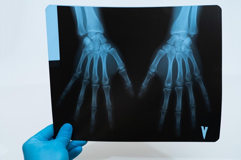
Radiological images allow doctors to identify specific features associated with psoriatic arthritis. These include bone erosions (areas where bone has been damaged or worn away), joint space narrowing (indicating cartilage loss between bones), and new bone formation at sites where tendons attach to bones.
The use of radiology in diagnosing and monitoring PsA allows for early detection and intervention when necessary. This early intervention can help prevent further damage to joints and improve patient outcomes overall.
So next time you visit your doctor for a check-up or evaluation related to your psoriatic arthritis diagnosis – don’t be surprised if they order some imaging tests! These invaluable tools help healthcare professionals gain a deeper understanding of your condition and guide appropriate treatment strategies
The Role of Radiology in Diagnosing and Monitoring Psoriatic Arthritis
Psoriatic arthritis is a complex condition that affects both the skin and joints. It can be challenging to diagnose, as its symptoms often overlap with other types of arthritis. This is where radiology plays a vital role in helping healthcare professionals accurately diagnose and monitor psoriatic arthritis.
Radiology, specifically imaging techniques such as X-rays, ultrasounds, and magnetic resonance imaging (MRI), allows doctors to visualize the internal structures of the body. By using these imaging techniques, they can detect any abnormalities or changes in the joints affected by psoriatic arthritis.
X-rays are commonly used in psoriatic arthritis radiology because they provide valuable information about joint damage and erosion. They help identify characteristic features such as bone loss, joint space narrowing, and bony proliferation known as “pencil-in-cup” deformities.
Ultrasound is another useful tool in diagnosing psoriatic arthritis. It can show inflammation around tendons and ligaments that may not be visible on an X-ray. Ultrasound also helps assess disease activity by detecting synovitis (inflammation of the lining of the joint) and enthesitis (inflammation at tendon attachments).
Magnetic resonance imaging (MRI) is particularly helpful for evaluating soft tissues like muscles, tendons, and ligaments affected by psoriatic arthritis. It provides detailed images that aid in assessing disease severity and identifying early signs of joint damage.
One advantage of radiological techniques is their ability to differentiate between different types of arthritis. For example, distinguishing between osteoarthritis (wear-and-tear) and psoriatic arthritis becomes possible through specific patterns seen on X-rays or MRI scans.
While radiology has proven invaluable in diagnosing psoriatic arthritis accurately; it does have some limitations. For instance,”silent” inflammation may not always appear on imaging tests despite ongoing disease activity experienced by patients.
Types of Imaging Used in Psoriatic Arthritis Radiology
When it comes to diagnosing and monitoring psoriatic arthritis, radiology plays a crucial role. By utilizing various imaging techniques, healthcare professionals can get a clearer picture of the affected joints and surrounding tissues. These images help in determining the presence and severity of inflammation, joint damage, and other characteristic features associated with this condition.
One commonly used imaging modality is X-ray, which provides detailed images of bones and joints. It can help identify hallmark signs of psoriatic arthritis such as erosions, bone proliferation, and joint space narrowing. X-rays are particularly useful for assessing structural changes over time.
Ultrasound is another valuable tool in psoriatic arthritis radiology. It allows for real-time visualization of soft tissues like tendons, ligaments, and synovium. This technique helps detect synovitis (inflammation of the lining around joints) which is a key finding in psoriatic arthritis.
Magnetic resonance imaging (MRI) offers a more comprehensive view by providing detailed images of bones, soft tissues, cartilage structures, as well as inflammatory changes within the joints. It can reveal early signs of disease activity before they become visible on x-rays.
Computed tomography (CT) scans may be used to evaluate bony anatomy when more precise details are required or if there are concerns about fractures or deformities.
These different types of imaging serve distinct purposes in the diagnosis and management process for psoriatic arthritis patients; however each has its own benefits and limitations that must be considered by healthcare providers when formulating treatment plans
Benefits and Limitations of Radiology in Psoriatic Arthritis
When it comes to diagnosing and monitoring psoriatic arthritis, radiology plays a crucial role. It provides valuable insights into the underlying disease processes, helping healthcare professionals make informed treatment decisions. Today, we will explore the benefits and limitations of radiology in managing psoriatic arthritis.
One of the key advantages of using radiology in psoriatic arthritis is its ability to detect early signs of joint damage. By capturing images of affected joints, such as X-rays or magnetic resonance imaging (MRI), doctors can identify structural changes before they become apparent clinically. This allows for timely intervention and potentially prevents further joint deterioration.
Furthermore, radiological imaging can help differentiate between different types of arthritis. For instance, by examining characteristic features on X-rays or MRI scans, physicians can distinguish between osteoarthritis and psoriatic arthritis accurately. This distinction is essential because each condition requires a tailored approach to treatment.
In addition to diagnosis, radiology aids in monitoring disease progression over time. Regular imaging assessments can track changes in joint inflammation or bone erosion patterns associated with psoriatic arthritis. These findings guide healthcare providers in adjusting treatment plans accordingly and assessing their effectiveness.
Common Findings on Radiological Images of Psoriatic Arthritis
Psoriatic arthritis, a chronic inflammatory condition that affects both the skin and joints, can be challenging to diagnose accurately. However, radiology plays a crucial role in identifying characteristic features and patterns associated with this condition.
When it comes to psoriatic arthritis radiology, several imaging techniques are commonly used. X-rays are often the initial choice as they can reveal changes in bone structure such as erosions and joint space narrowing. Ultrasound is another valuable tool that allows for real-time visualization of inflammation and fluid accumulation within the joints.
MRI scans provide detailed images of soft tissues like tendons, ligaments, and synovium. They help detect early signs of inflammation not visible on x-rays or ultrasound. Additionally, CT scans may be used to assess bone density and identify enthesitis (inflammation at sites where tendons attach to bones).
These imaging modalities aid in differentiating between psoriatic arthritis and other conditions like osteoarthritis. One key difference is the presence of erosions at joint margins seen more frequently in psoriatic arthritis patients compared to those with osteoarthritis.
Furthermore, radiological images also help healthcare professionals assess disease progression over time. They track changes in bone density loss or new erosions which indicate worsening disease activity.
Radiology plays a vital role in diagnosing psoriatic arthritis by identifying characteristic findings on various imaging techniques such as x-rays, ultrasound, MRI scans, and CT scans. These findings assist clinicians in distinguishing between different types of arthritis while also aiding treatment planning based on disease severity assessment through monitoring changes seen over time on subsequent images obtained during follow-up visits!
How Radiology Aids in Treatment Planning for Psoriatic Arthritis
Radiology plays a crucial role in the treatment planning for psoriatic arthritis. By providing detailed images of the affected joints and surrounding tissues, radiologists can help rheumatologists make informed decisions about appropriate treatment options.
One way that radiology aids in treatment planning is by assessing disease activity and severity. Through imaging techniques such as X-rays, ultrasound, and magnetic resonance imaging (MRI), doctors can evaluate the extent of joint damage and inflammation. This information helps determine whether aggressive treatments like biologic medications are necessary or if milder interventions may be sufficient.
Furthermore, radiological findings can guide targeted therapy approaches. For example, imaging may reveal specific patterns of joint involvement or characteristic features associated with psoriatic arthritis. Understanding these patterns can help clinicians devise individualized treatment plans tailored to each patient’s needs.
In addition to aiding in diagnosis and guiding treatment decisions, radiology also contributes to monitoring disease progression over time. Regular imaging assessments allow doctors to track changes in joint damage or inflammation levels. This enables them to adjust medication dosages or switch therapies if needed to ensure optimal outcomes for patients.
By providing valuable insights into disease activity, severity, and response to treatment, radiology significantly enhances the ability of healthcare providers to plan effective strategies for managing psoriatic arthritis. With ongoing advancements in technology and image analysis techniques, this field continues evolving to better serve patients living with this chronic condition.
Conclusion
Radiology plays a crucial role in diagnosing and monitoring psoriatic arthritis. By utilizing various imaging techniques, healthcare professionals can identify key features and patterns that are indicative of this condition. The use of radiology not only helps differentiate psoriatic arthritis from other types of arthritis, such as osteoarthritis, but also aids in treatment planning.
Through radiological imaging, doctors can assess the severity of joint damage and inflammation, allowing for personalized treatment plans to be developed. This leads to better management of symptoms and improved quality of life for individuals living with psoriatic arthritis.
While radiology is a valuable tool in the diagnosis and monitoring of psoriatic arthritis, it does have its limitations. It cannot provide a definitive diagnosis on its own; rather, it complements other clinical assessments and tests. Additionally, interpretation of radiological images requires expertise to accurately identify specific findings associated with psoriatic arthritis.
Despite these limitations, there is no denying the significant contribution that radiology makes in managing this complex disease. With ongoing advancements in technology and research, we can expect even greater insights into the pathophysiology and progression of psoriatic arthritis through radiological studies.
If you suspect you may have psoriatic arthritis or are already diagnosed with the condition, consult with your healthcare provider about incorporating radiological imaging into your treatment plan. Together with their expertise and the information provided by diagnostic imaging tools like X-rays or MRI scans, they can work towards finding effective strategies to alleviate pain and manage symptoms associated with this chronic inflammatory disease.


