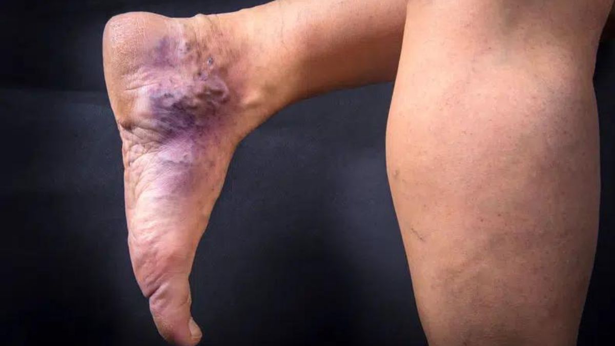
Accumulation of iron in tissues causes a characteristic reddish discoloration known as hemosiderin staining, often called hemosiderosis or iron staining. It typically manifests as in spots with a history of bleeding or blood vessel injury. Staining with hemosiderin is not limited to any one organ and usually indicates a more serious underlying health issue. The purpose of this page is to provide a synopsis of hemosiderin staining, including its characteristics, causes, diagnostic criteria, and therapeutic alternatives.
Causes of Hemosiderin Staining
Hemosiderin staining results from the release of iron from dead red blood cells. Its growth may be influenced by a number of factors:
- Chronic Venous Insufficiency: Chronic venous insufficiency (CVI) is a leading cause of hemosiderin staining. When blood flow in the veins of the legs is impaired, chronic venous insufficiency (CVI) develops. Hemosiderin deposition occurs when blood pools in the legs for an extended period of time, causing inflammation and red blood cell leaking.
- Trauma: Hemosiderin staining can occur after any major injury or trauma that causes bleeding. This can be the result of anything from a routine operation to a broken bone.
- Hemorrhagic Disorders: Hemosiderin staining can be caused by conditions that increase the risk of bleeding, like hemophilia or von Willebrand disease.
- Vascular Disorders: Hemosiderin staining occurs when blood flow is impeded and blood cells leak out, as in the case of varicose veins, venous stasis ulcers, or deep vein thrombosis.
Diagnosis of Hemosiderin Staining
Medical history, a physical exam, and diagnostic testing are often used together to make a diagnosis of hemosiderin staining. In order to determine the root of the problem, a doctor will examine the affected area and review the patient’s medical history. Tests that can help in diagnosis are:
- Dermoscopy: In dermoscopy, a handheld instrument is used to look at the top layer of skin. It’s useful for identifying the severity of hemosiderin staining and distinguishing it from other skin disorders.
- Biopsy: It may be necessary to remove a small piece of the diseased tissue and examine it closely under the microscope. Hemosiderin deposits can be identified and other possible causes eliminated.
- Doppler Ultrasound: Doppler ultrasound is frequently utilized for the purpose of evaluating blood flow and identifying venous insufficiency or other vascular problems.
Treatment Options
Hemo’siderin staining treatment centers on resolving the underlying cause and alleviating associated symptoms. The following strategies could be applied:
- Compression Therapy: Compression stockings or bandages may be prescribed for patients with chronic venous insufficiency to enhance blood flow and alleviate edema.
- Wound Care: Vein stasis ulcers require special attention to the wound in order to heal and avoid infection. Cleaning, dressing, and applying topical treatments are all possibilities.
- Surgical Interventions: Vein stripping, endovenous ablation, or sclerotherapy are a few examples of surgical treatments used to treat venous insufficiency and stop additional hemo’siderin staining.
- Dermatological Treatments: Hemo’siderin staining can be treated using dermatological procedures such laser therapy, chemical peels, and microdermabrasion. These methods can bleach the damaged skin, making the discoloration less noticeable.
Here’s more information on hemosiderin staining
- Hemo’siderin staining occurs as a result of the breakdown of red blood cells and the release of iron. When red blood cells are damaged or destroyed, the iron they contain is released and taken up by cells called macrophages. Macrophages are responsible for recycling iron and storing it as hemosiderin, a complex form of iron.
- The accumulation of hemosiderin leads to the characteristic brownish discoloration seen in tissues. This staining can occur in various organs and tissues, including the skin, lungs, liver, spleen, and kidneys. However, it is most commonly observed in the skin, particularly in areas that have experienced previous bleeding or trauma.
- Chronic venous insufficiency (CVI) is one of the primary causes of hemosiderin staining. CVI occurs when the valves in the veins of the legs fail to function properly, leading to poor blood flow and increased pressure in the veins. This can result in the leakage of red blood cells and hemosiderin deposition in the surrounding tissues.
- Trauma or injury that involves bleeding can also lead to hemo’siderin staining. Surgeries, fractures, or any significant tissue damage that involves bleeding can cause the release of red blood cells and subsequent accumulation of hemosiderin in the affected area.
- Hemorrhagic disorders, such as hemophilia or von Willebrand disease, can increase the risk of bleeding and contribute to hemosiderin staining. These conditions impair the blood’s ability to clot effectively, resulting in prolonged bleeding and subsequent hemosiderin deposition.
- Certain vascular disorders can also cause hemosiderin staining. Varicose veins, which are enlarged and twisted veins, can lead to blood pooling and leakage, resulting in hemosiderin accumulation. Venous stasis ulcers, which are open sores that develop due to poor blood flow, can also contribute to hemosiderin staining.
- The diagnosis of hemosiderin staining involves a thorough evaluation by a healthcare professional. They will consider the patient’s medical history and perform a physical examination to identify potential underlying causes. Dermoscopy, which involves using a handheld device to examine the skin’s surface, can help differentiate hemo’siderin staining from other skin conditions and determine the extent of the staining. In some cases, a biopsy of the affected tissue may be performed to confirm the presence of hemosiderin deposits and rule out other potential causes.
- The treatment of hemosiderin staining aims to address the underlying cause and manage symptoms. For cases related to chronic venous insufficiency, compression therapy is often recommended. Compression stockings or bandages can help improve blood circulation, reduce swelling, and prevent further hemosiderin deposition. Proper wound care is essential for venous stasis ulcers, including regular cleansing, appropriate dressings, and the use of topical medications.
- In some situations, surgical interventions may be necessary to treat the underlying venous insufficiency and prevent further hemo’siderin staining. Procedures such as vein stripping, endovenous ablation, or sclerotherapy can help improve blood flow and reduce the accumulation of hemosiderin.
- Dermatological treatments may be utilized to improve the appearance of hemo’siderin staining. Laser therapy, chemical peels, or microdermabrasion can help lighten the affected skin and reduce the visibility of the discoloration.
Conclusion
Hemo’siderin staining, in conclusion, is an iron deposition in tissues that causes a reddish discoloration called hemosiderin staining. Vascular disease, trauma, hemorrhagic diseases, and chronic venous insufficiency are common causes. In order to make a diagnosis, doctors need to look at patient histories, examine them, and sometimes do tests. Compression therapy, wound care, surgical procedures, and dermatological therapies are all used to treat the underlying cause and alleviate symptoms.


